Tag Archives: diffusion tensor imaging

Women’s Multitasking Skill Linked to Neural Network Patterns

Diane Halpern

Diane Halpern

Differences between men’s and women’s performance on cognitive tasks, particularly mathematics and science have been observed for decades, with men generally excelling at motor and spatial tasks and women excelling in memory and social cognition tasks.

Camilla Benbow

Camilla Benbow

Claremont McKenna College’s Diane F. Halpern led an extensive review of these performance differences with Camilla P. Benbow of Vanderbilt University, University of Missouri‘s David C. Geary, Ruben C. Gur of University of Pennsylvania, Janet Shibley Hyde and Morton Ann Gernsbacher of University of Wisconsin. 

David Geary

David Geary

Their evidence “provided no single or simple answer” to contrasting skills by gender but a comprehensive brain imaging study of more than 400 males and more than 500 females between ages 8 and 22 years, provides evidence for popular observations.

Madhura Ingalhalika

Madhura Ingalhalika

Using diffusion tensor imaging, University of Pennsylvania’s Madhura Ingalhalikar, Alex Smith, Drew Parker, Theodore D. Satterthwaite, Mark A. Elliott, Kosha Ruparel, Raquel E. Gur, Ruben C. Gur and Ragini Verma with Hakon Hakonarson of Children’s Hospital of Philadelphia, demonstrated that male and female brains differ in the network of neural connections.
Known as the “structural connectome,” these connections between neural structures were described by Indiana University’s Olaf Sporns, who reviewed imaging techniques to visualize their activity.

Ted Satterthwaite

Ted Satterthwaite

These gender-linked structural differences result in differing competencies.
Ingalhalikar’s team observed that male brains structures show more connections within the front and back of the brain hemisphere in the supratentorial region.

Olaf Sporns

Olaf Sporns

This area connects perception and coordinated action and enables males’ skill in quickly perceiving and applying information to a single complex task, spatial reasoning, and learning motor skills.

Ingalhalikar connectome
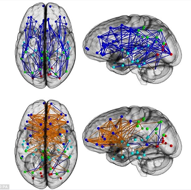
In contrast, female brains contain more neural connections between hemispheres in supratentorial regions. 
This connection pattern enables females to recall faces and execute multiple complex tasks simultaneously more easily than males due the increased neural connections between analytical and intuitive processing modes.

Dardo Tomasi

Dardo Tomasi

Building on earlier work on these differences by Brookhaven National Lab’s Dardo Tomasi and Nora D. Volkow of National Institute on Drug Abuse, Ingalhalikar’s team found these differences were reversed in the cerebellar connections, where male brains showed greater intrahemispheric connectivity and female brains demonstrated more interhemispheric connections.

Nora Volkow

Nora Volkow

These structural differences lead to different development for girls and boys from an early age, and result in significant, less modifiable differences by adolescence and adulthood. 

Frequently-observed differences in male and female performance are rooted in different neural connection patterns by gender.

 -*What exceptions have you seen to findings of women’s skill in multitasking and social insight, and men’s competence in spatial reasoning and motor skill acquisition?

RELATED POSTS:

Follow-share-like http://www.kathrynwelds.com and @kathrynwelds

Twitter  @kathrynwelds
Blog – Kathryn Welds | Curated Research and Commentary  
Google+
LinkedIn Open Group Psychology in Human Resources (Organisational Psychology)
Facebook Notes

©Kathryn Welds

Advertisement

Juggling as Brain Training

The physical skill of juggling can change the brain’s structure and function for the better, and may be a recommended therapy for brain injuries.

Bogdan Draganski

Bogdan Draganski

Volker Busch

Volker Busch

Bogdan Draganski, now of University of Lausanne collaborated with University of Regensburg colleagues Volker Busch, Ulrich Bogdahn, and Gerhard Schuierer, and Arne May, now of University of Hamburg  with Christian Gaser of University of Jena, to visualize brain plasticity among volunteers who learned to juggle.

Ulrich Bogdahn

Ulrich Bogdahn

Volunteers showed transient and selective structural changes in the left parietal lobe’s posterior cortex  and bilateral central temporal areas brain, areas associated with processing and storing complex visual motion on used whole-brain magnetic-resonance imaging.

Gerhard Schuierer

Gerhard Schuierer

This study demonstrates that the brain’s macroscopic structure can change based on stimuli like juggling, rather than being limited to functional changes in the cortex.

Arne May

Arne May

May and Gaser collaborated with University of Hamburg colleagues Janina Boyke, Joenna Driemeyer, and Christian Büchel in related research.

Christian Gaser

Christian Gaser

This time, they trained 25 people with an average age of 60 years in juggling for 12 weeks, and 25 control group volunteers were not trained.

Christian Büchel

Christian Büchel

The team conducted three MRI brain scans for each participant:

  • Before juggling practice
  • After 3 months of juggling
  • After another 3 months of no juggling.

Jugglers showed significant increase in the brain’s “gray matter,” nerve cells’ bodies responsible for information processing, located in the hippocampus (memory formation), bilateral nucleus accumbens (reward systems that may lead to action) and visual cortext’s middle temporal area.

Without practice during the three months after the training, none of the volunteers retained their ability to juggle and their gray matter declined to pre-training levels.

This suggests the value of continued practice in physical and cognitive skills to maintain brain structure and function.

Jan Scholz

Jan Scholz

University of Oxford’s Jan Scholz, Miriam Klein-FlüggeTimothy E.J. Behrens, and Heidi Johansen-Berg extended this research to demonstrate that people who learn to juggle also increased “white matter” containing axons that connect different cells, not just gray matter.

 The Oxford team conducted baseline brain scans using diffusion tensor imaging to reveal  white matter structure for 24 young men and women volunteers, who later practiced juggling for half an hour a day for six weeks.

Miriam Klein-Flügge

Miriam Klein-Flügge

The researchers compared brain scans of 24 non-juggling volunteers and found that the volunteer jugglers increased white matter in the parietal lobe’s intraparietal sulcus, which integrates vision and reaching and grasping in the periphery of vision.
Even less-skilled jugglers had similar increases in white matter, attributed to amount of time devoted to practice.

Heidi Johansen-Berg

Heidi Johansen-Berg

In contrast, the non-jugglers showed no changes in white matter after the six week period, suggesting value in practicing juggling to develop the brain’s structure and functioning to enable rapid, coordinated movement and body positioning.

Brain scans taken after four weeks without juggling practice showed that the new white matter remained and the amount of gray matter increased, showing some skill retention in the absence of consistent practice.

-*What physical skills do you develop as brain training?

RELATED POSTS:

Twitter:    @kathrynwelds
Blog: Kathryn Welds | Curated Research and Commentary 
Google+:
LinkedIn Open Group Psychology in Human Resources (Organisational Psychology)
Facebook Notes:

©Kathryn Welds